
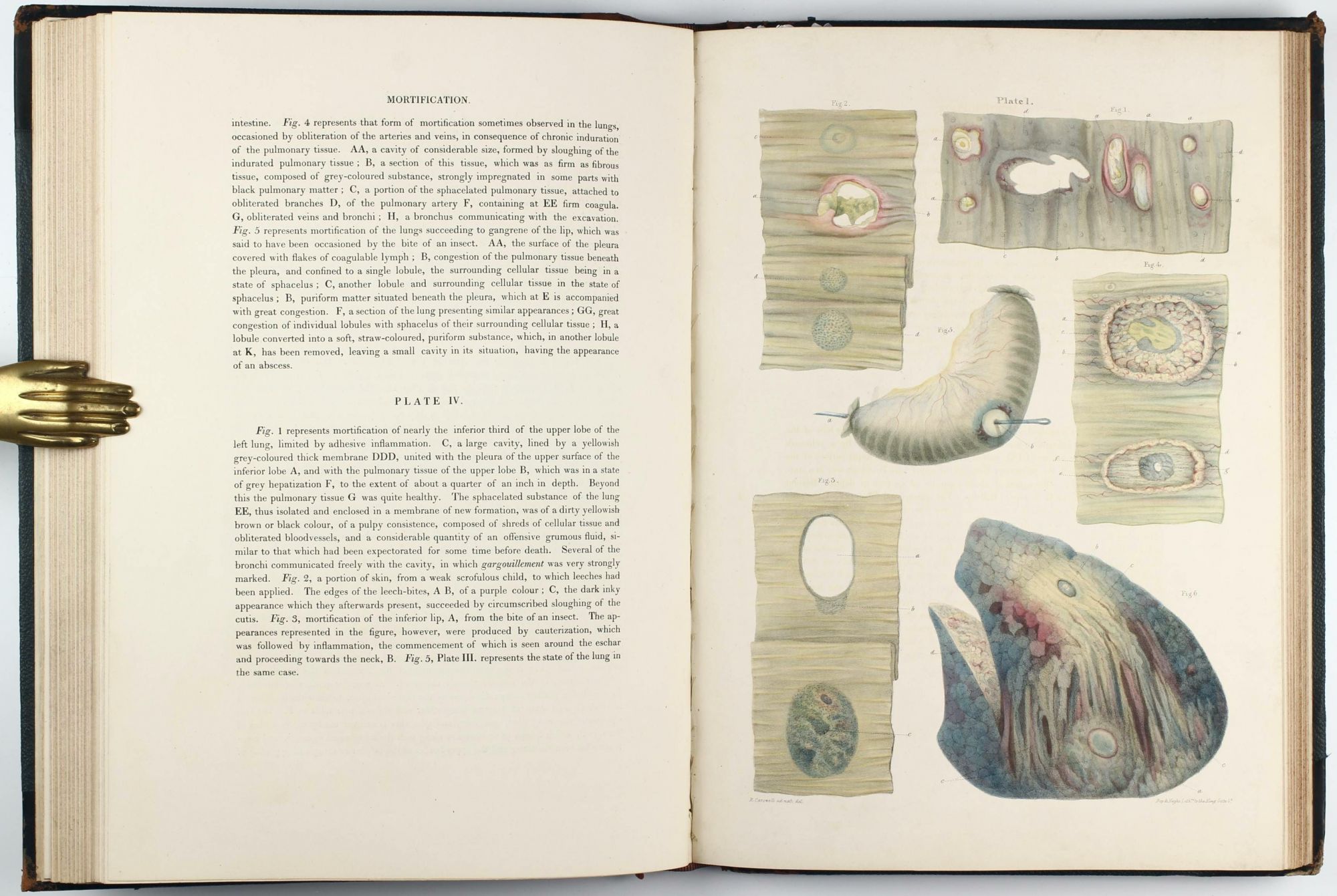
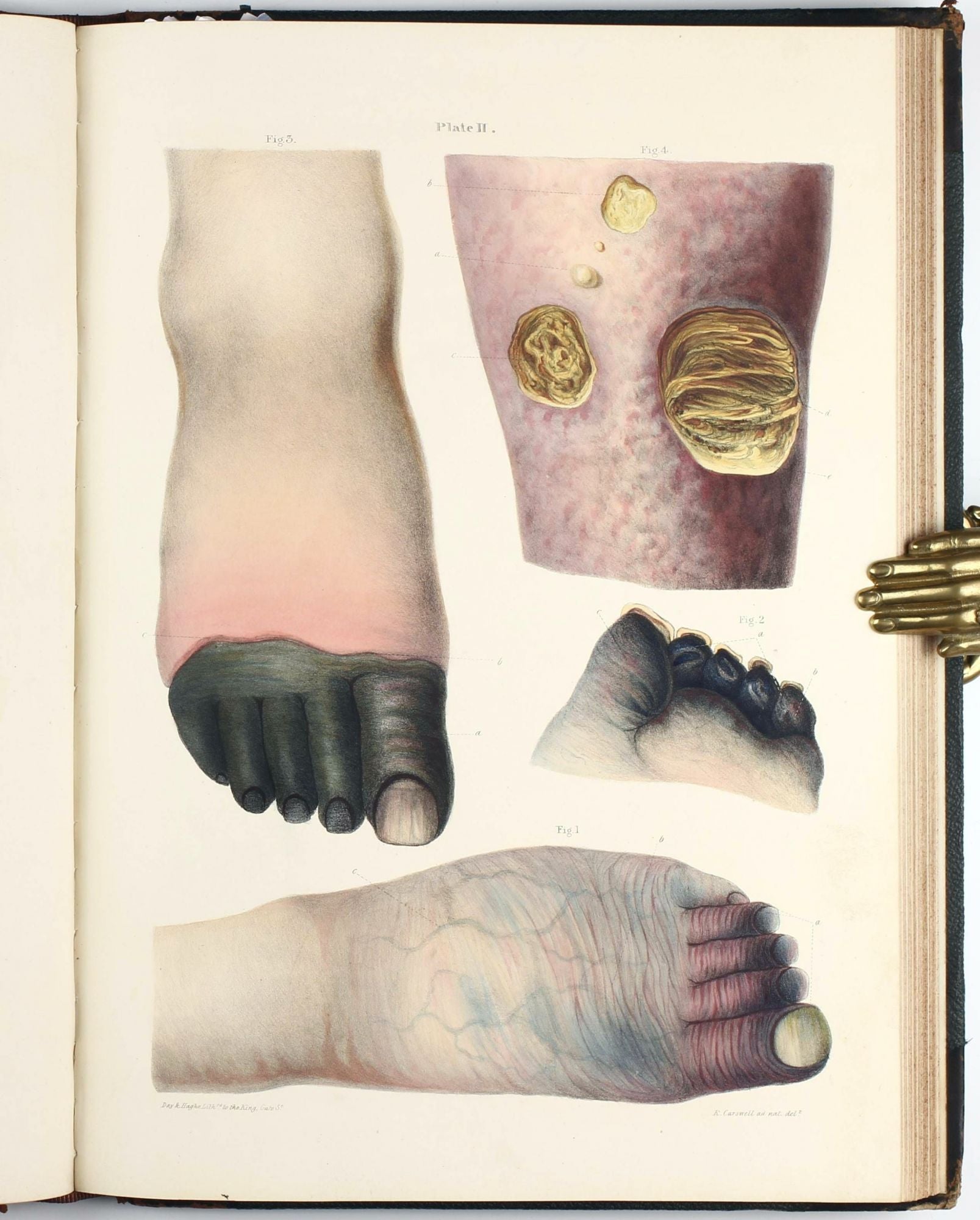
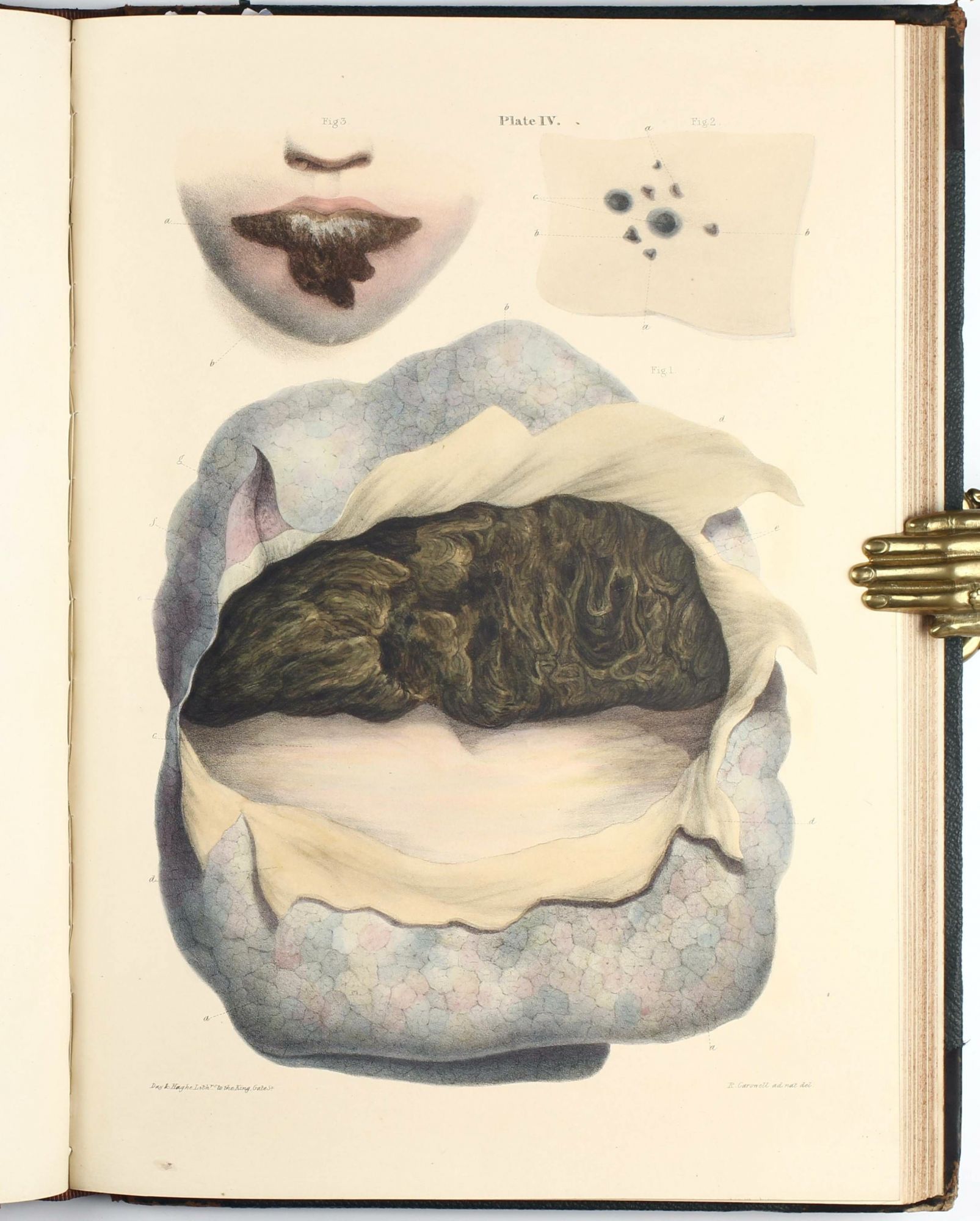
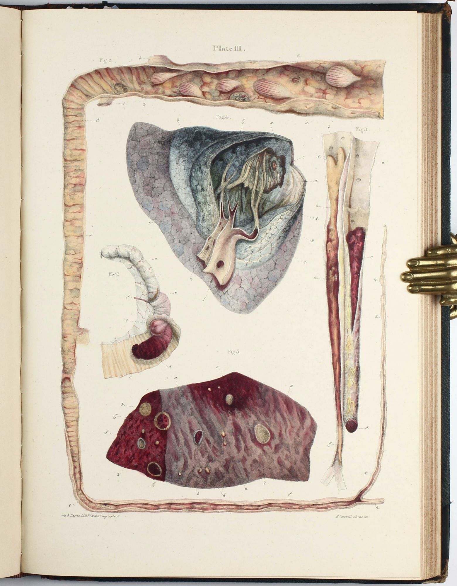
One of the most beautiful atlases of pathology
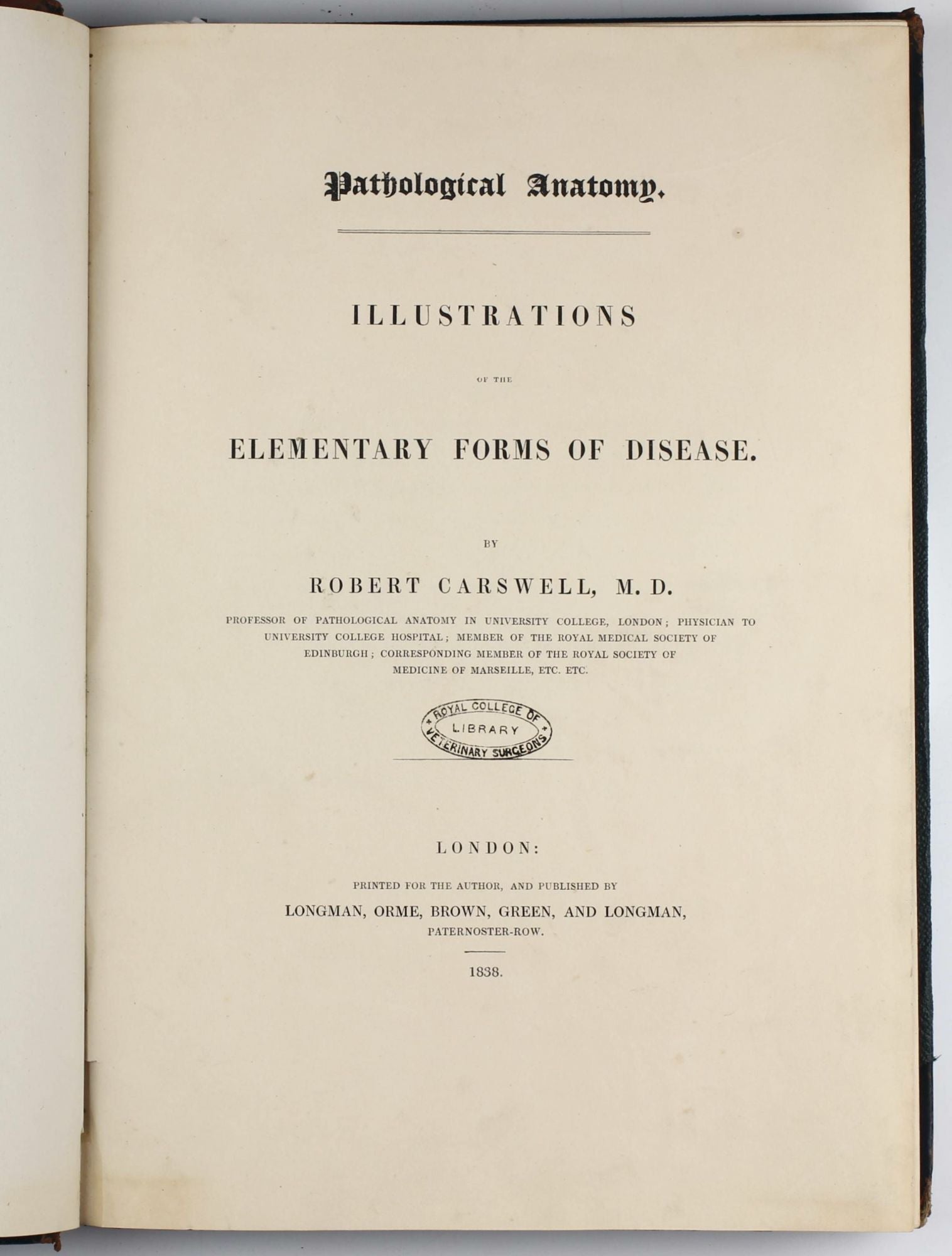
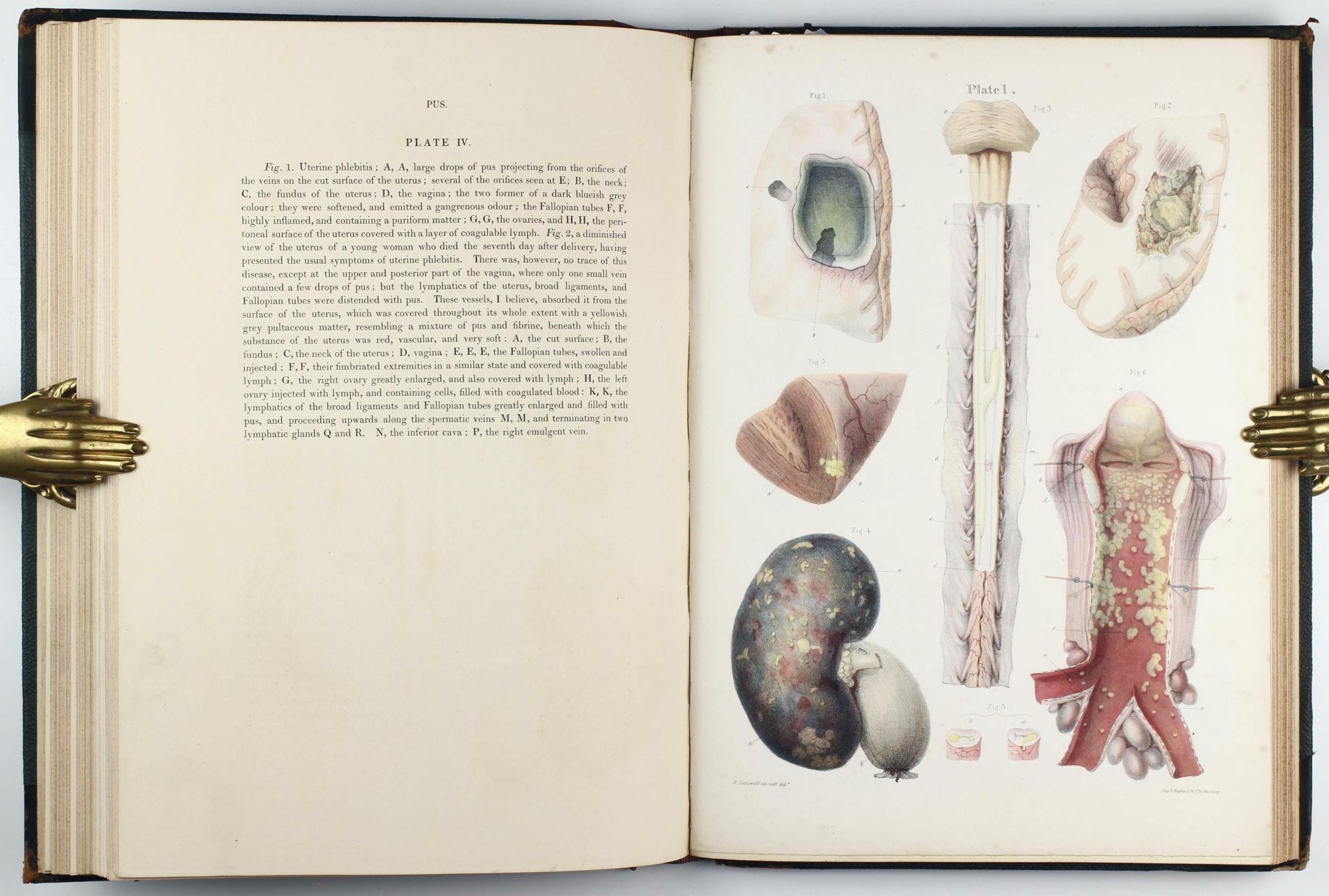
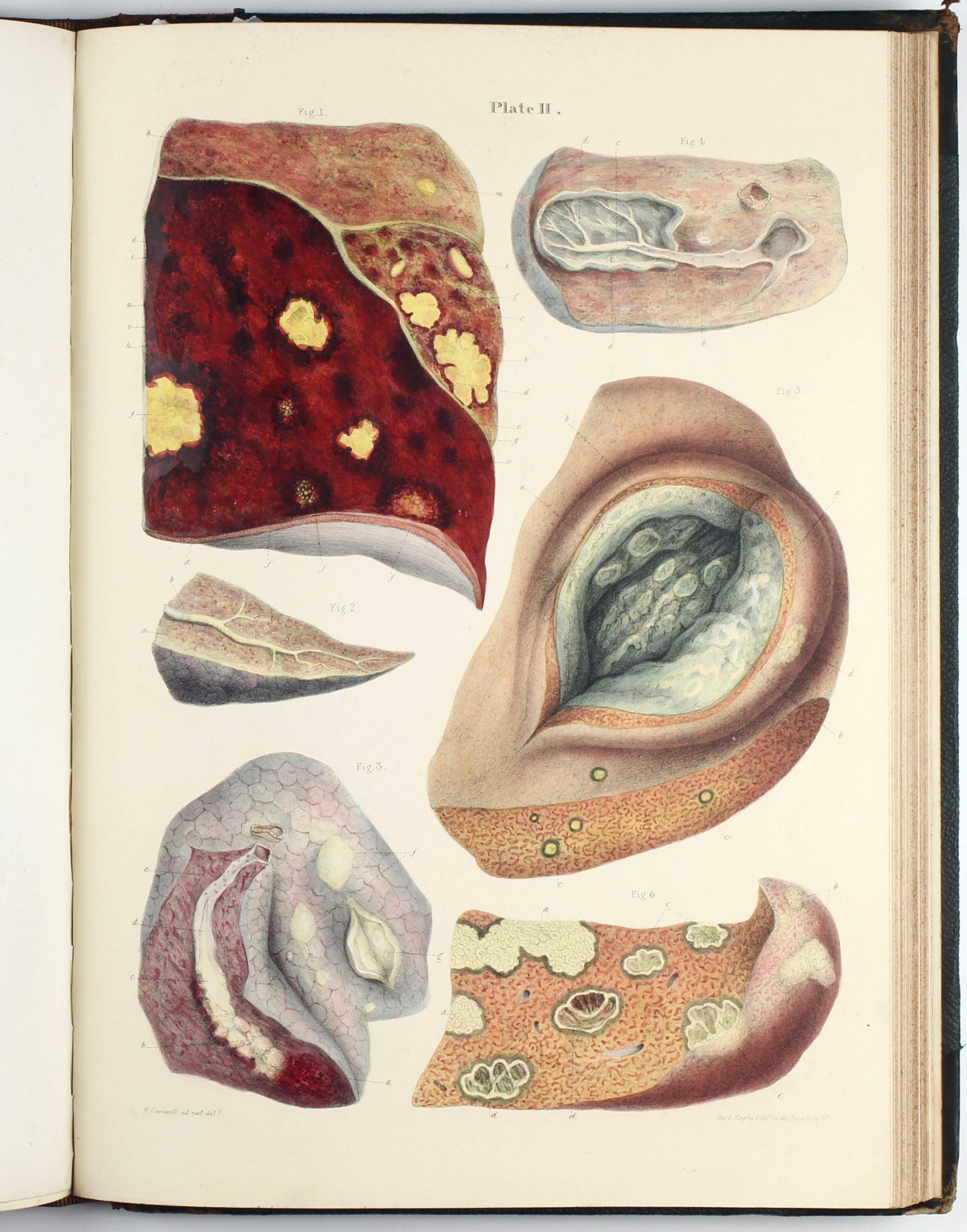
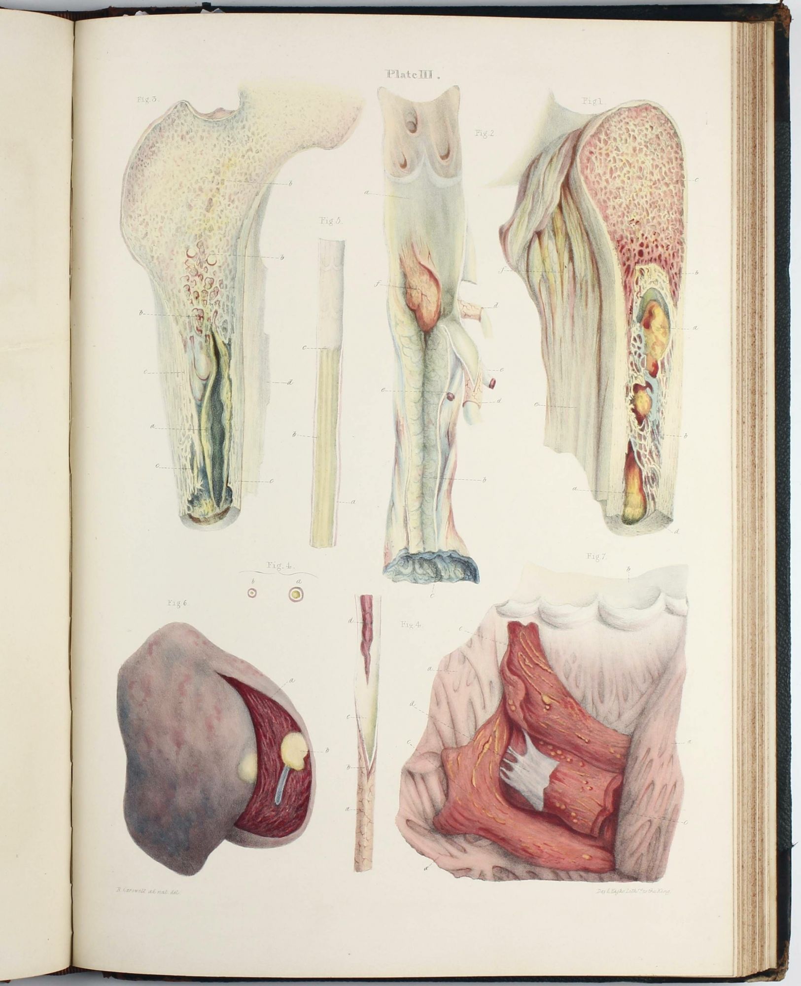
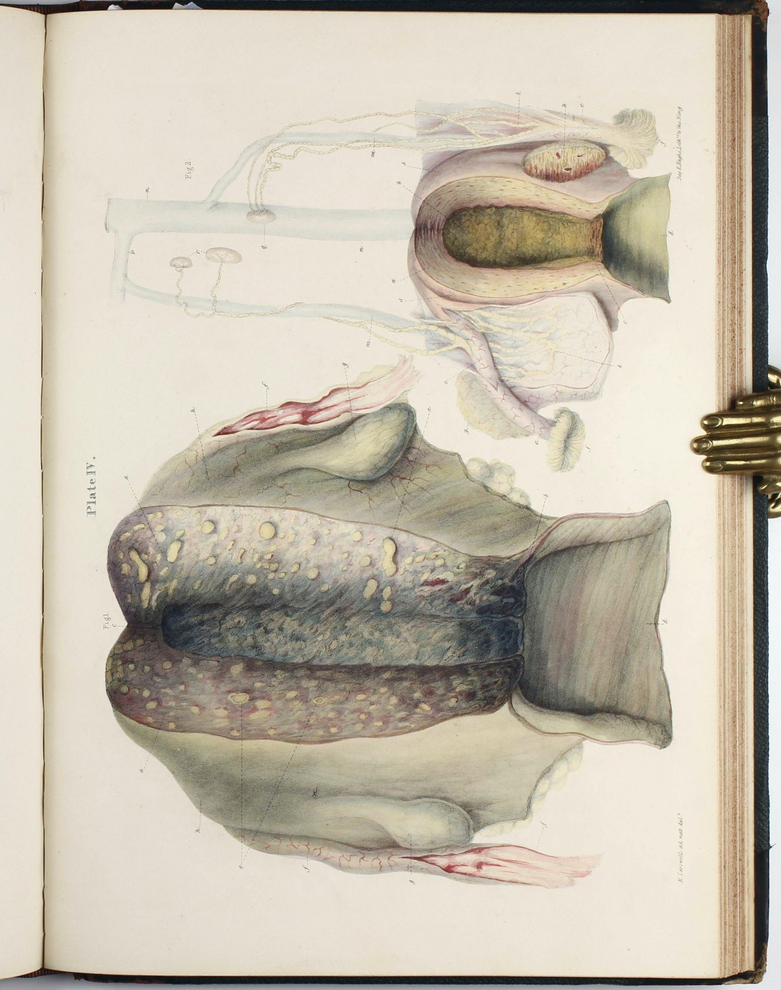
Pathological Anatomy. Illustrations of the Elementary Forms of Diseases.
London: Longman, Orme, Brown, Green and Longman for the author, 1838.
1st Edition. Hardcover. Very Good. Item #003269
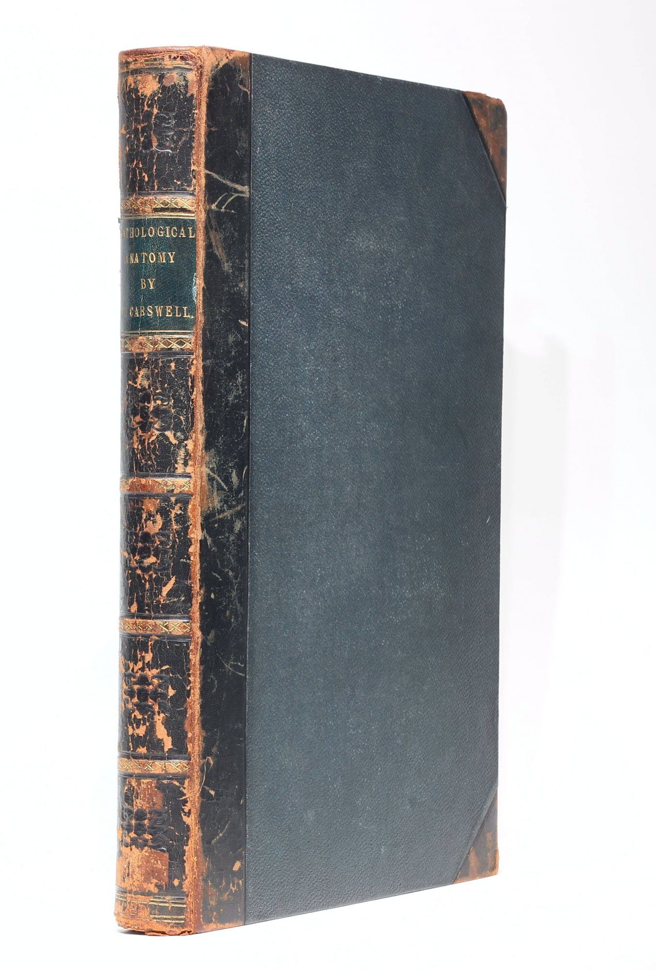
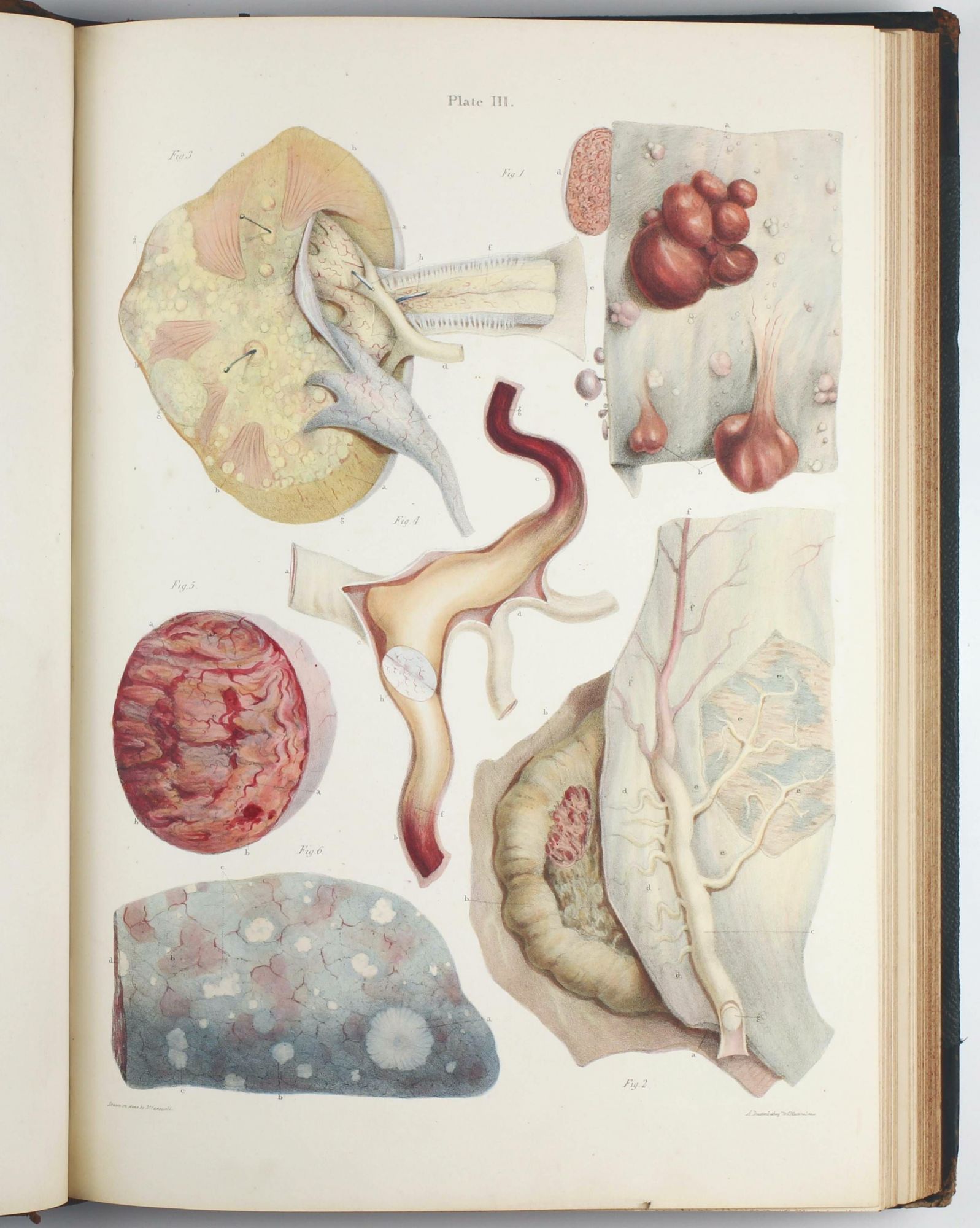
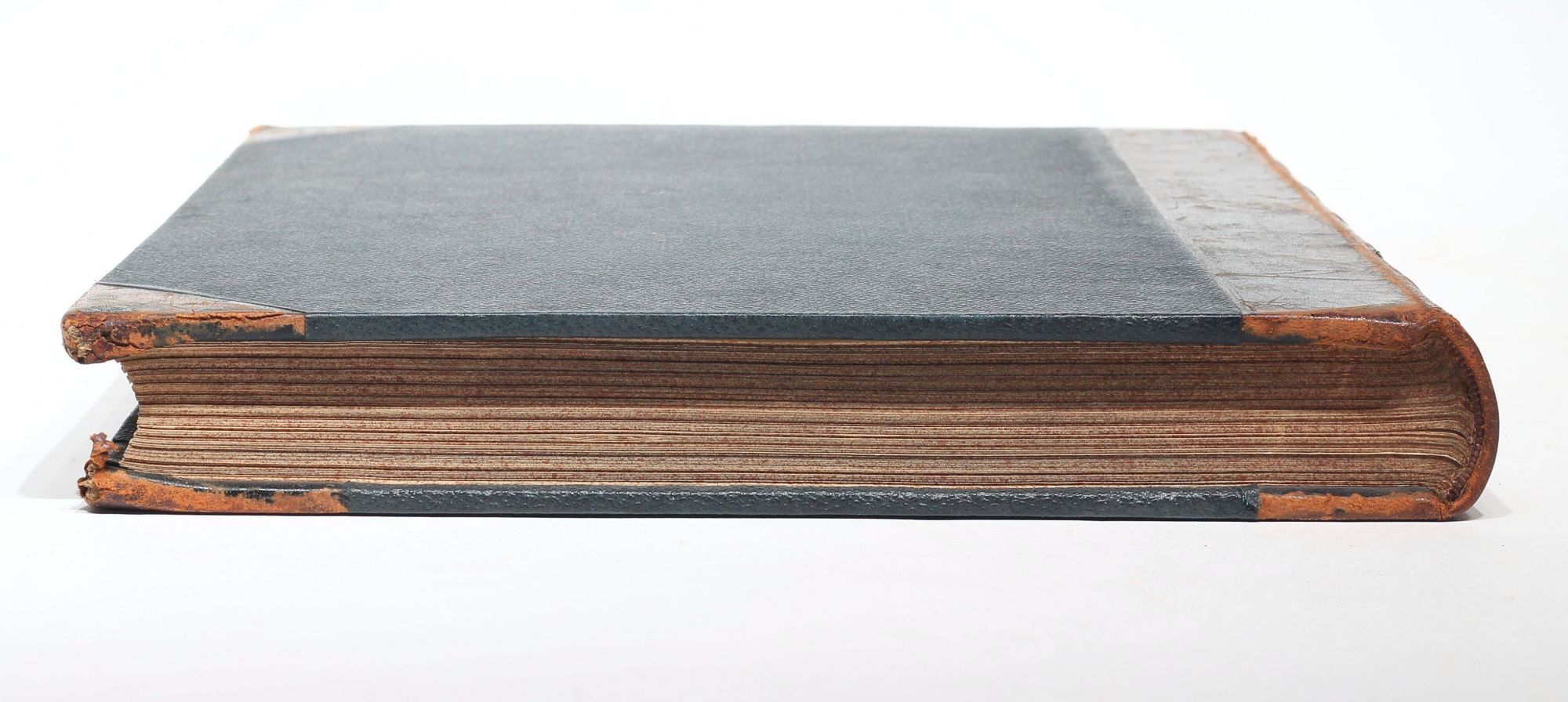
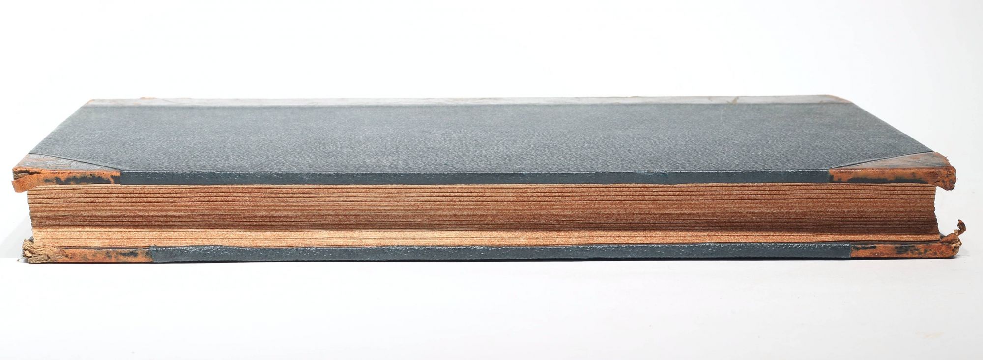
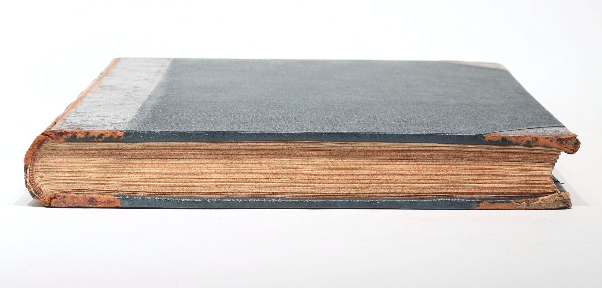
Folio (374 x 267 mm). 109 unnumbered leaves, 48 fine hand-colored lithographed plates after Carswell. Contemporary half calf over boards, spine with green morocco label lettered in gilt, some gilt decoration and blind stamping, red-sprinkled edges (spine leather and hinges heavily rubbed, wear to spine ends and corners). Internally very crisp and clean with little age-toning of text and plates only, two plates with offsetting from dark colors, minor spotting of one plate, rare marginal thumb-soiling. Provenance: Royal College of Veterinary Surgeons, London (ink stamp on title and a few other leaves); small sticker of bindery S. Weston, London to rear pastedown. Excellent copy. ----
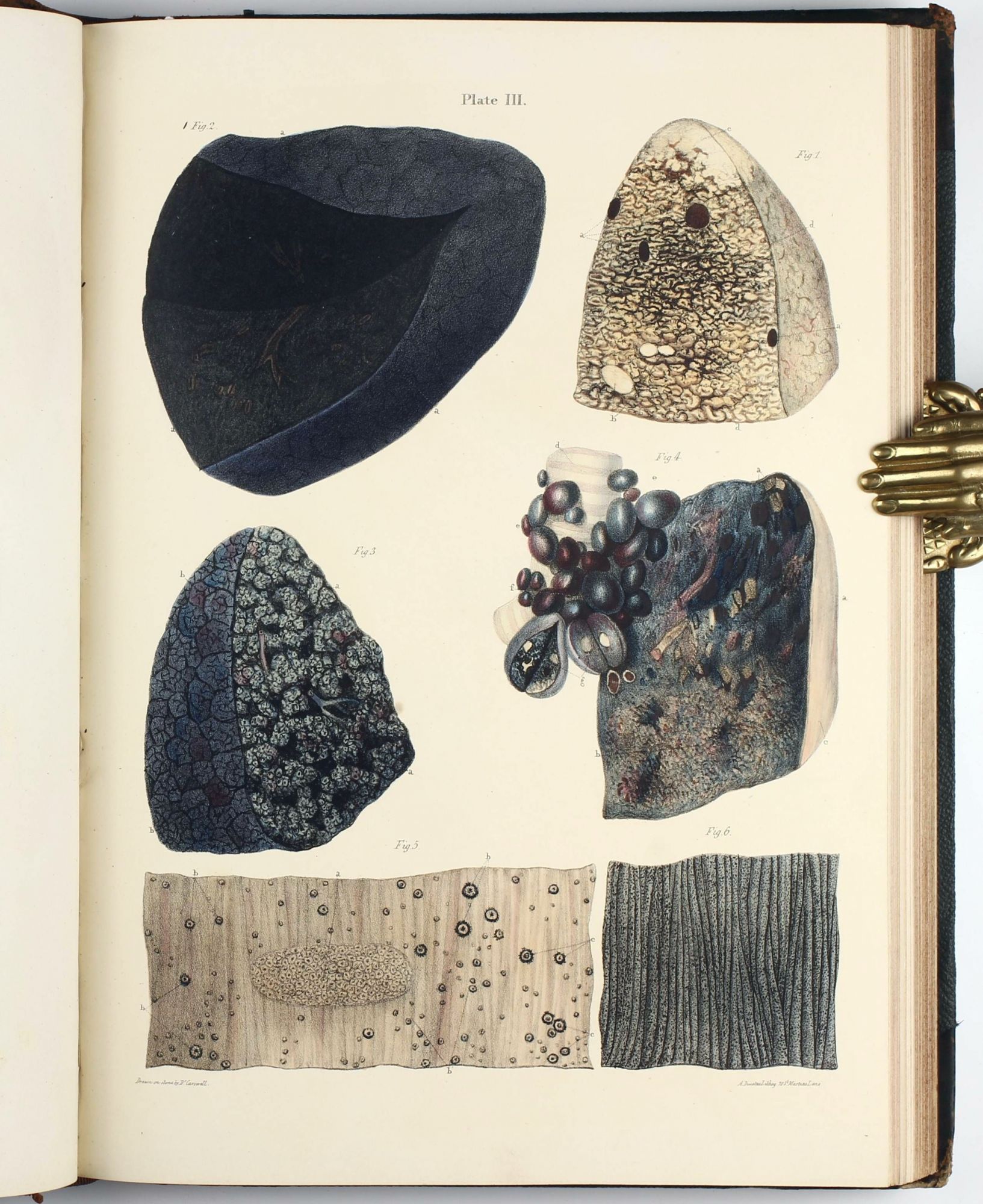
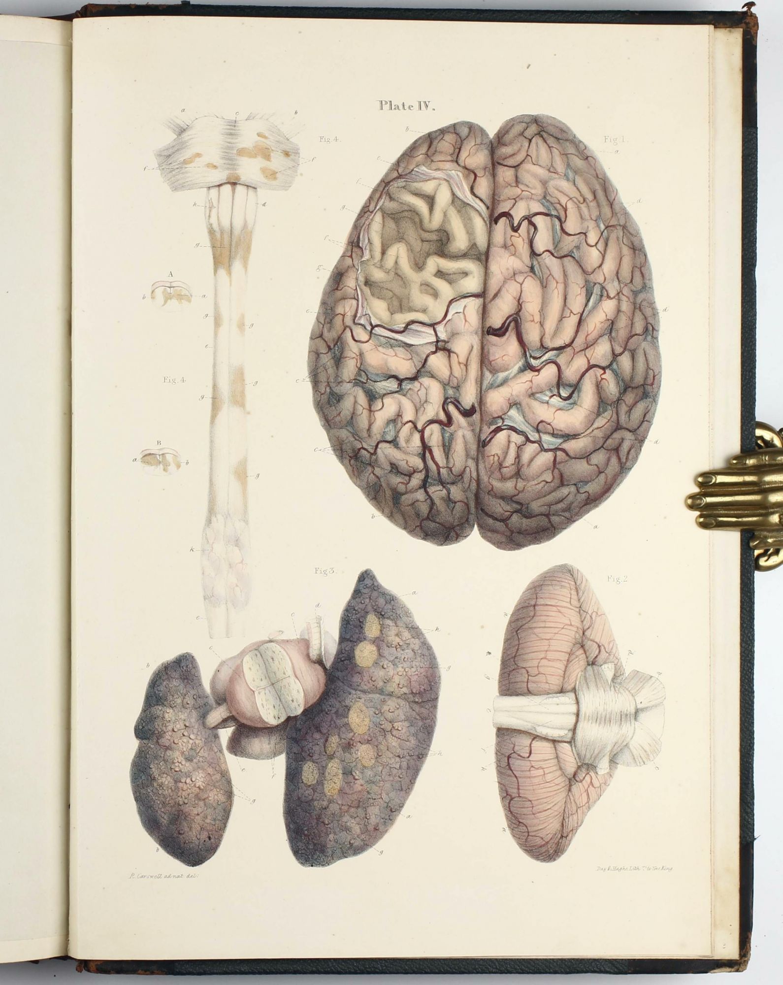
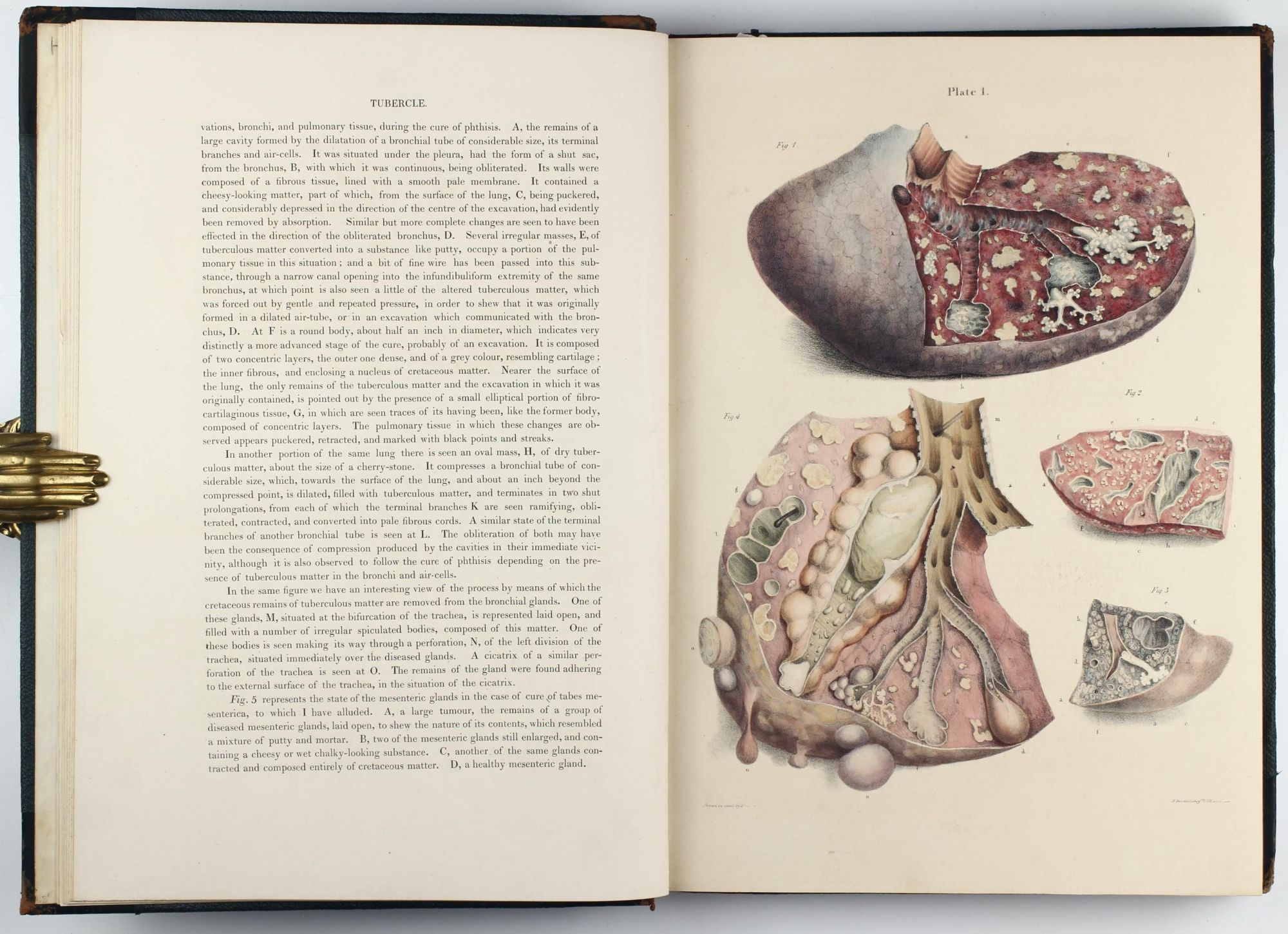
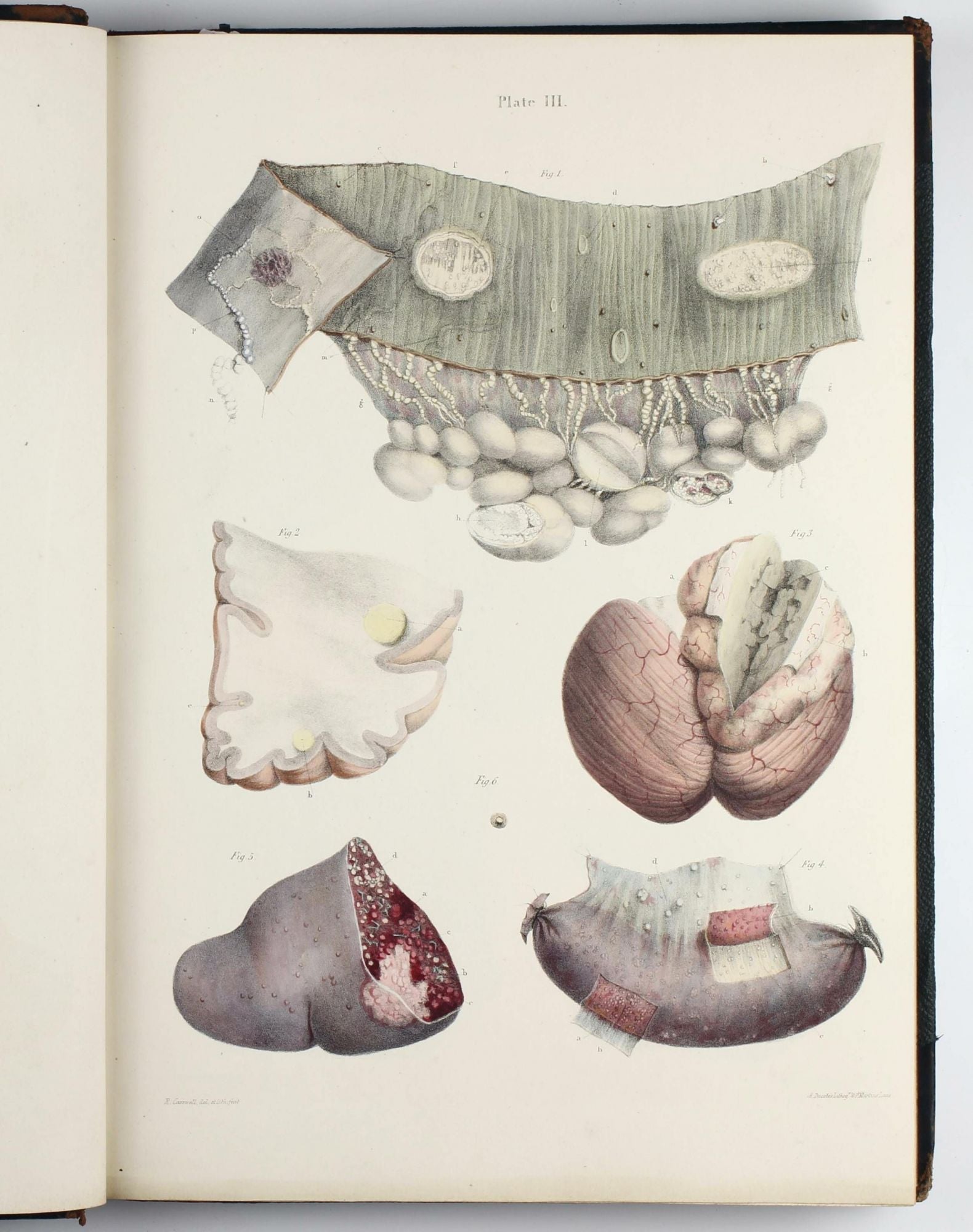
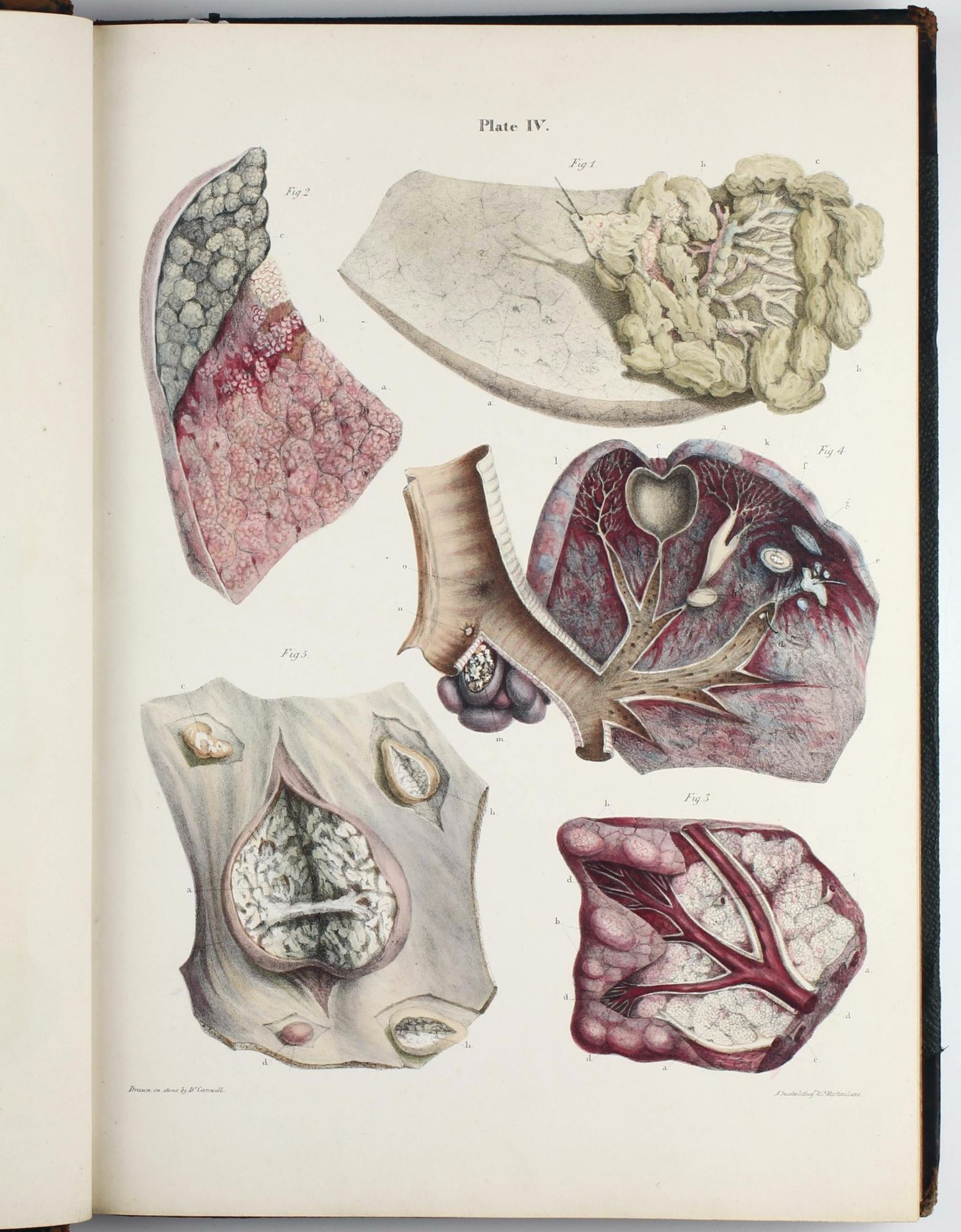
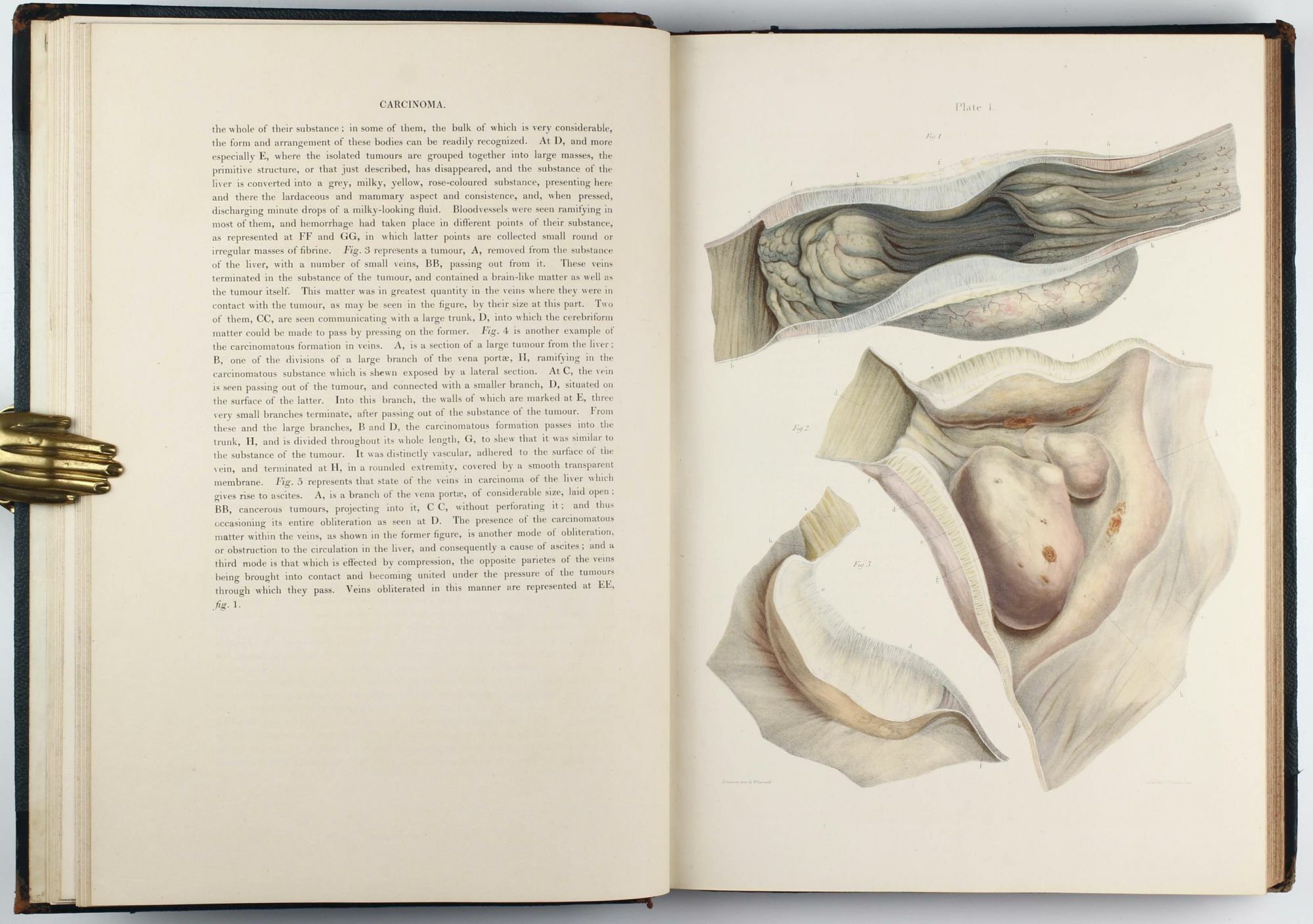
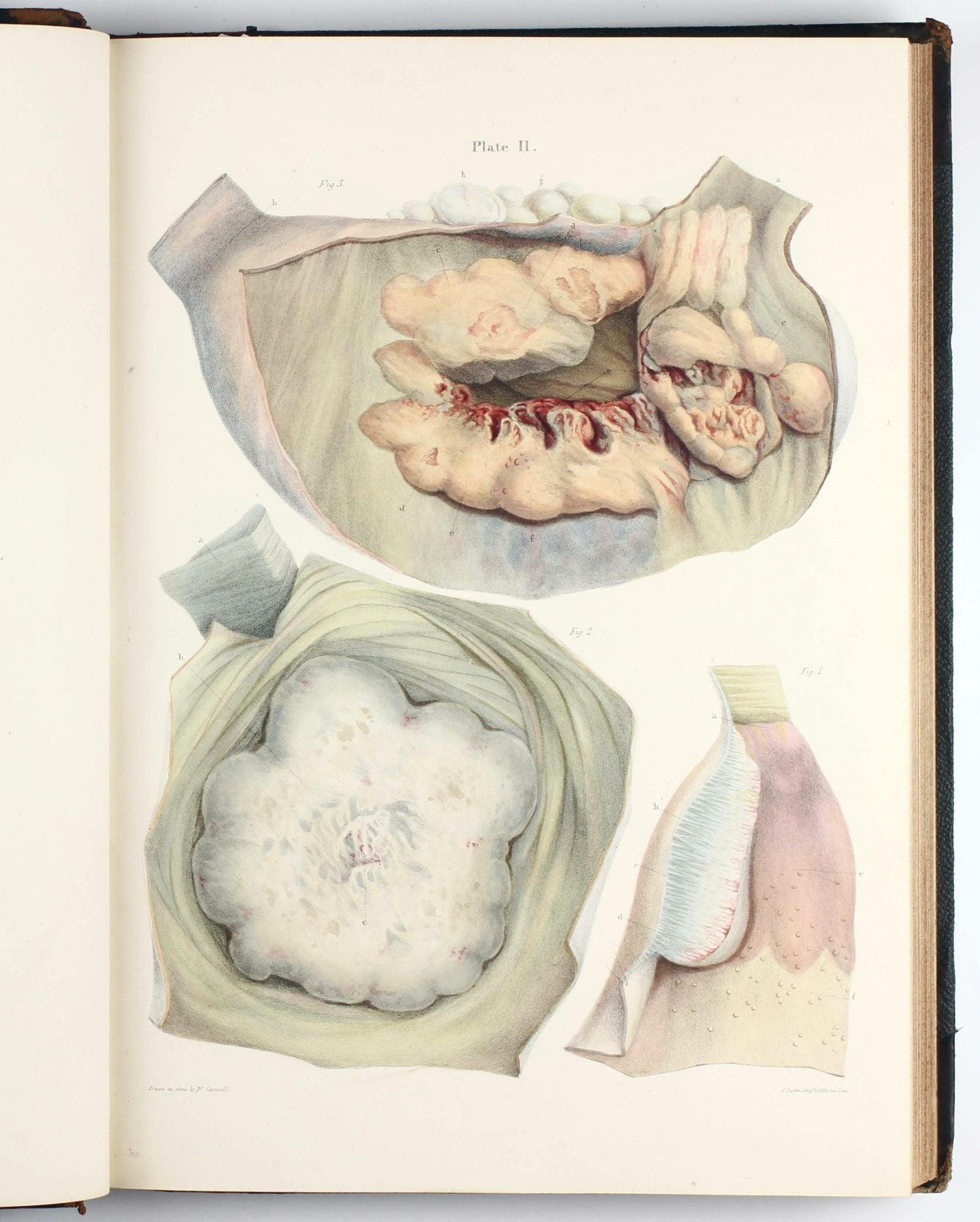
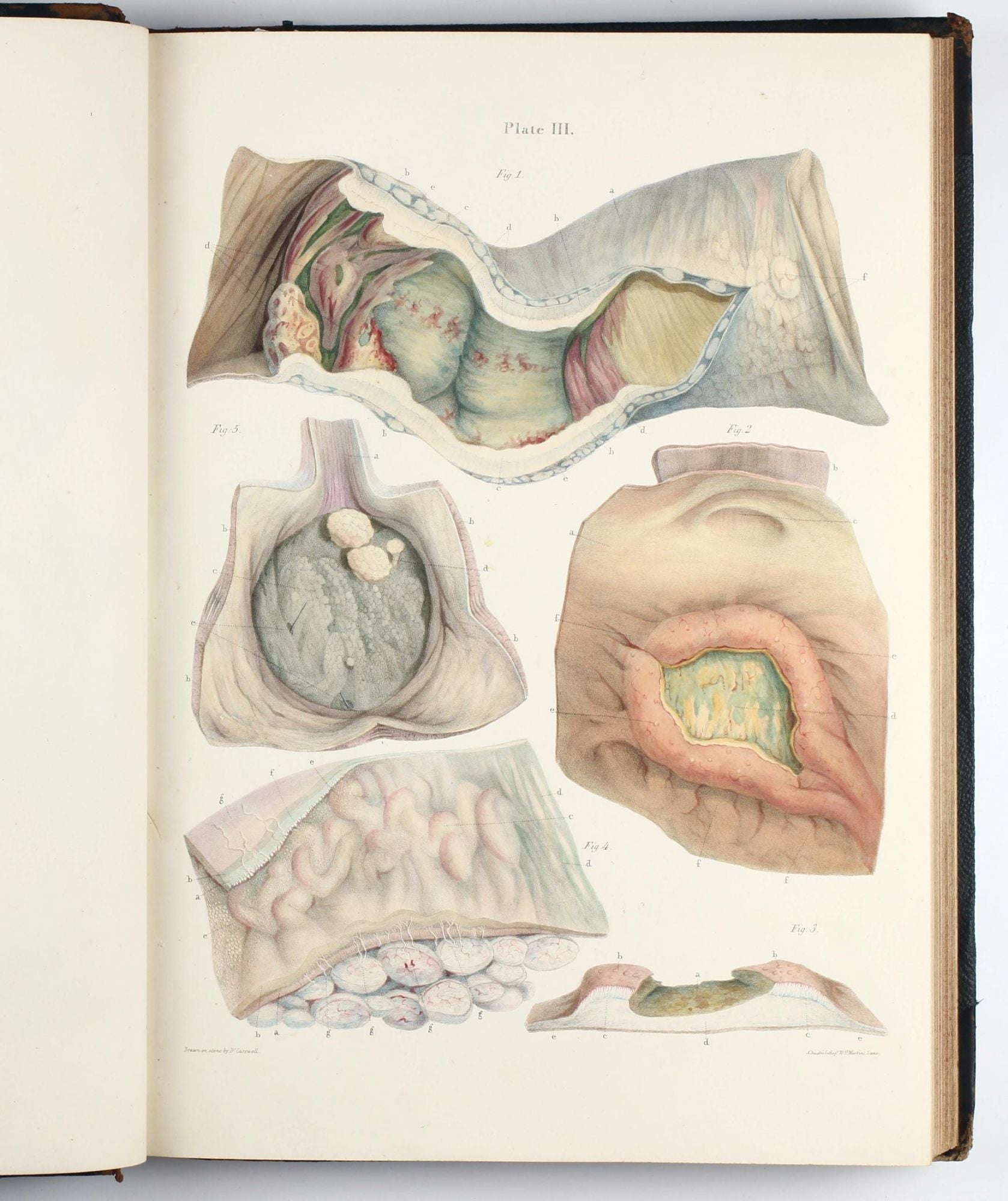
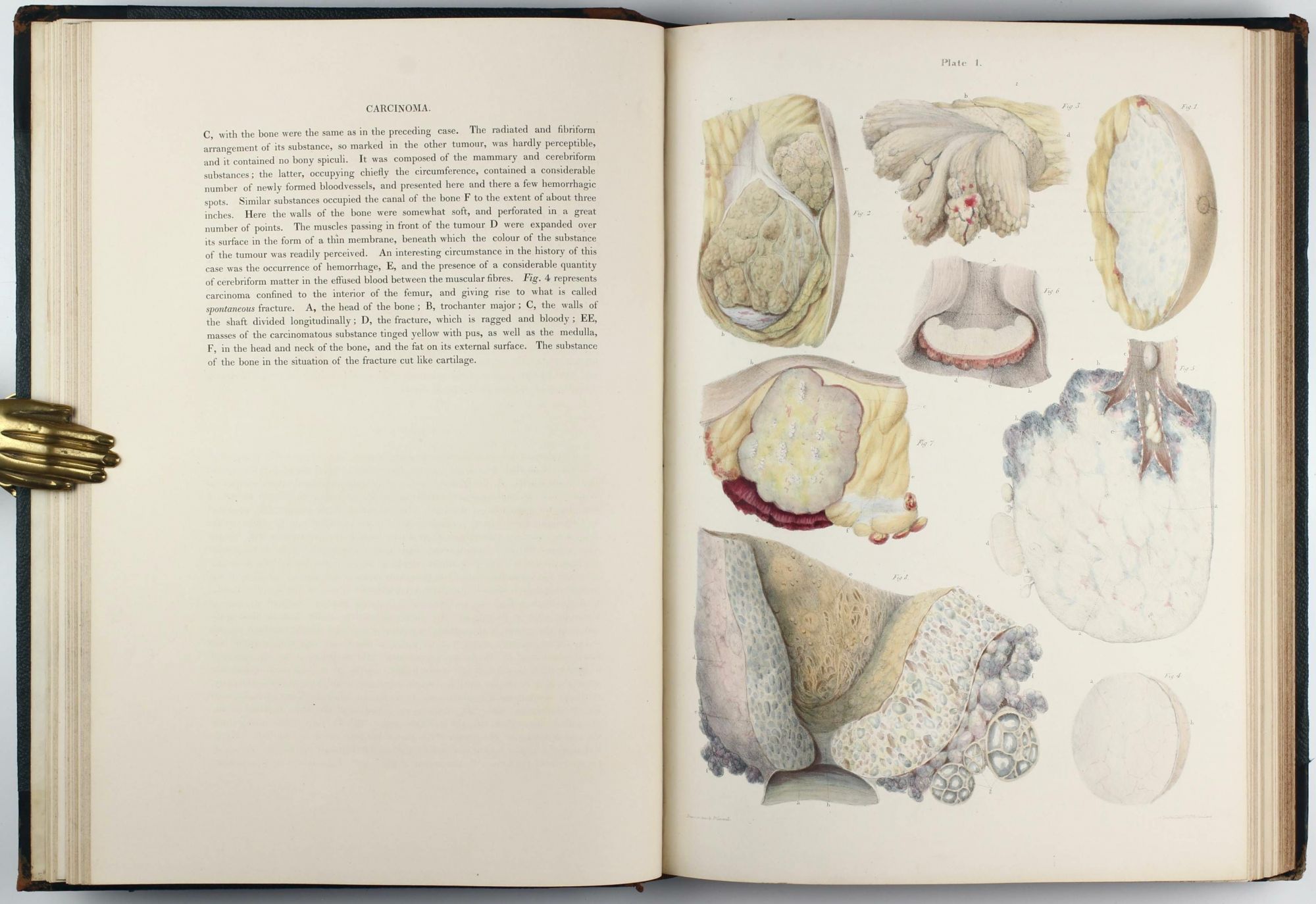
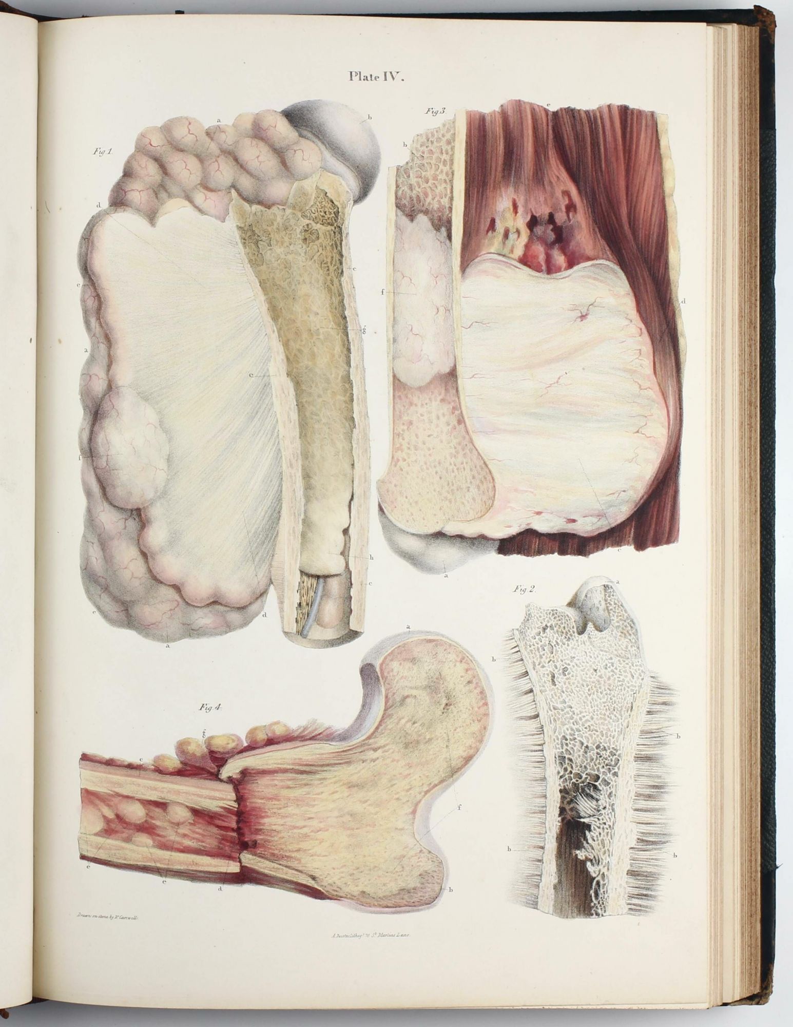
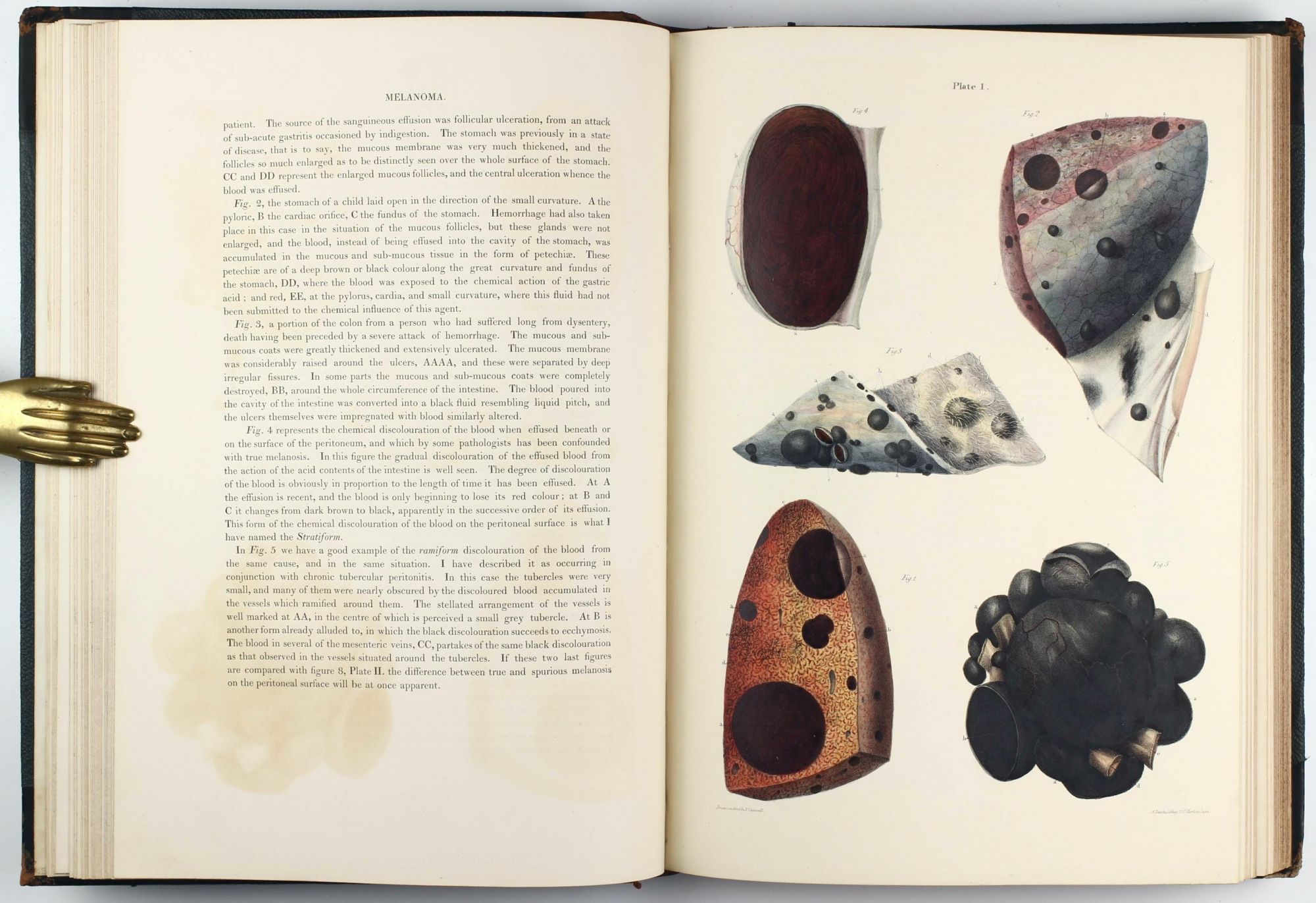
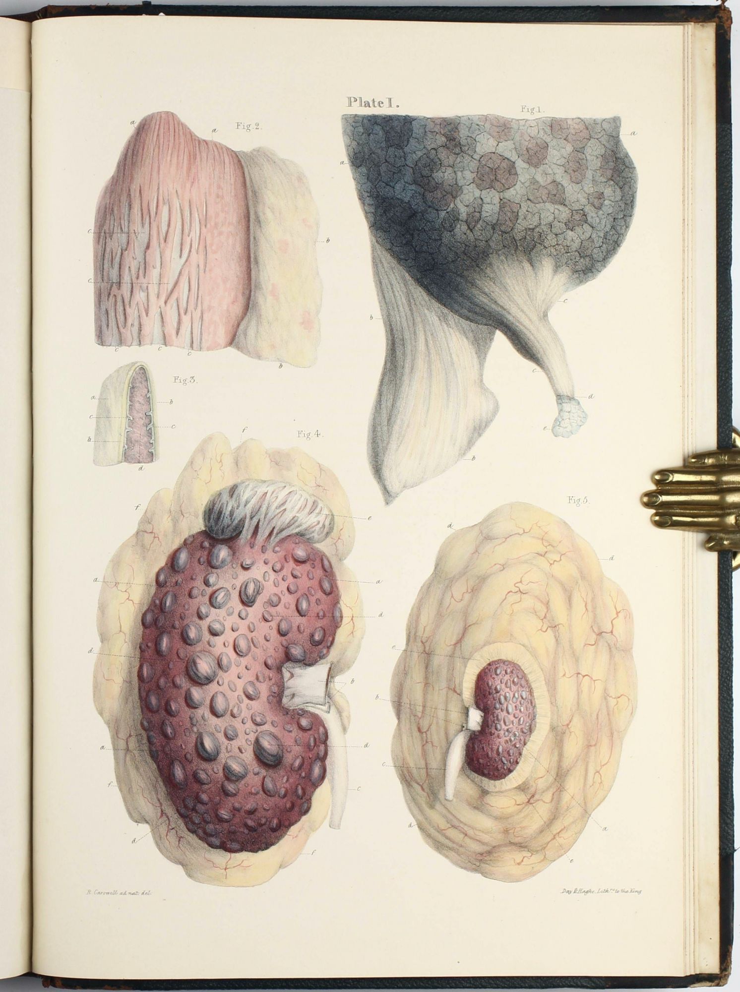
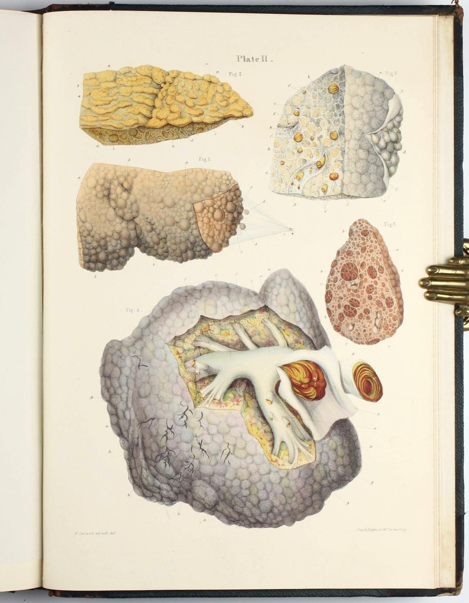
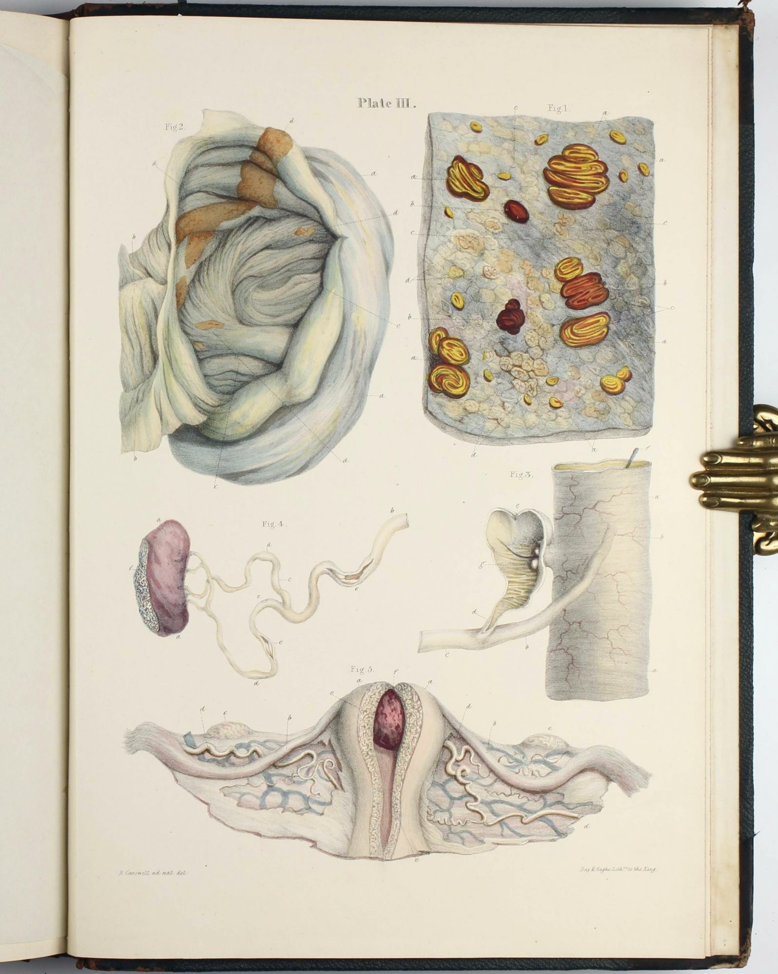
Eimas, Heirs of Hippocrates 1501; Norman 408; Osler 2250; Garrison-Morton 2291; Goldschmid 156; Wellcome II, p.306. - FIRST AND ONLY EDITION. Carswell's atlas, which he illustrated himself, is one of the most beautiful atlases of pathology. The work was "originally published in twelve Fascicles, beginning in January 1833 and continuing until January 1838 ... The Longman archive [. . .] records that between 1833 and 1840, 2361 fascicles (nos. 1-12) were sold; from this number we can infer that the first edition of Pathological anatomy was probably not greater than 300 copies." (Norman). The work's importance was swiftly recognized, and it remained esteemed throughout the century, as J. F. Payne's opinion of 1886 demonstrates: "These illustrations have, for artistic merit and for fidelity, never been surpassed, while the matter represents the highest point which the science of morbid anatomy had reached before the introduction of the microscope" (DNB).
Sir William Osler, who began his career as a pathologist, notes: "Carswell... studied morbid anatomy in Paris under Louis. He was commissioned by University College, London, to prepare a collection of pathological drawings, and in about three years (1828-31) he completed a series of 2,000 water-color drawings of diseased structures, which is still preserved at the College, where he was appointed professor of anatomy. The plates for his great work on pathological anatomy were furnished from his own drawings and put upon the stone by himself. These illustrations have, for artistic merit and for fidelity, never been surpassed, while the matter represents the highest point which the science of morbid anatomy had reached before the introduction of the microscope" (Osler, Bibliotheca Osleriana, 2250).
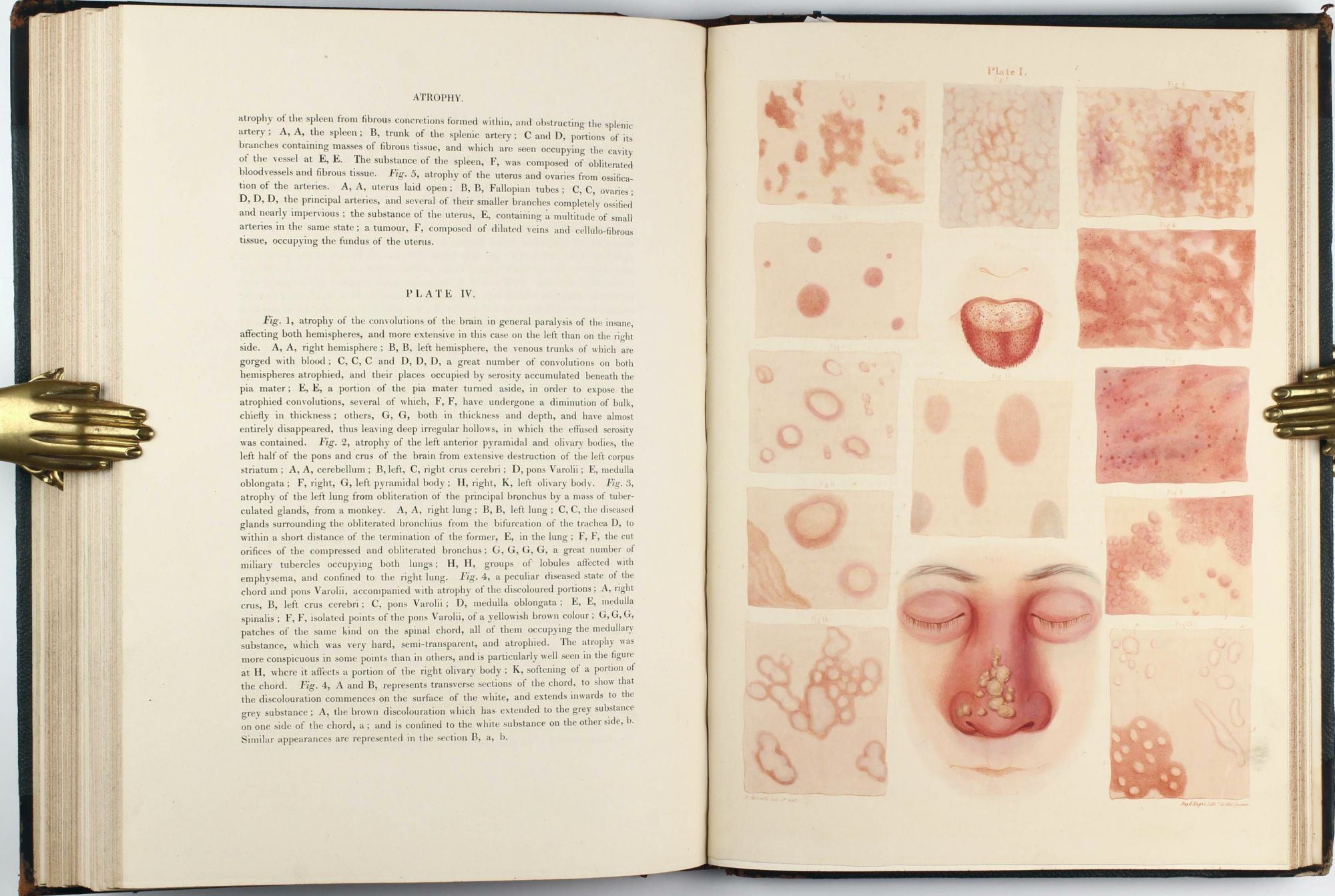
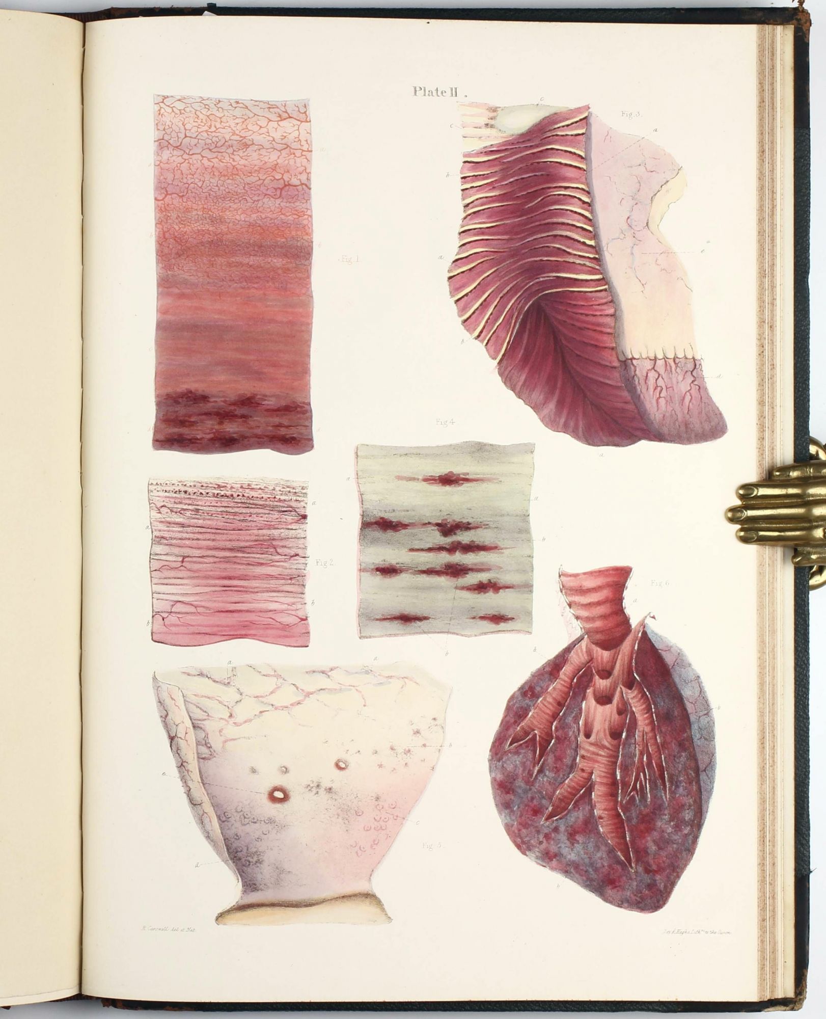
Rivaled for beauty and accuracy only by Cruveilhier's Anatomie pathologique du corps humain, this work "is rightly regarded as one of the finest pathological atlases ever produced." (Eimas, Heirs of Hippocrates 1501). "The beautiful hand-colored lithographed plates ... include good representations of post-mortem digestion of the stomach, cirrhosis of the liver, dry gangrene of the toes, endocarditis, and tuberculosis of the lungs and intestine" (Norman); these plates also include "the first illustration (in color) 'of the brain in general paralysis of the insane'" (Hunter and MacAlpine, Three Hundred Years of Psychiatry, 1535-1860, p. 784). - Visit our website to see more images!
Price: 22,000 € * convert currency
Delivery time up to 10 days. For calculation of the latest delivery date, follow the link: Delivery times
Lieferzeit max. 10 Tage. Zur Berechnung des spätesten Liefertermins siehe hier: Lieferzeiten